

Many youngsters who saw Jurassic Park wanted to get involved with dinosaurs and were inspired to become scientists. Dr. Michael PITTMAN, Assistant Professor at The Chinese University of Hong Kong (CUHK)’s School of Life Sciences had his interest sparked at a young age by Sir David Attenborough’s documentaries and became a palaeobiologist. Whatever his inspiration, he is the only palaeontologist in the world who co-developed a totally new imaging technique, laser stimulated fluorescence imaging (LSF), for research into fossils. He is now using this technology to walk a dual path in palaeontology and archaeology.
“My main focus in palaeontology is on the origin of flight but I also have a strong interest in archaeology. However, that is not the only area we are working on.”
A whole new light fluorescing fossils
In the last few months, the results of new significant palaeontology research from CUHK has been published by Dr. Pittman. Success in these studies came from the ground-breaking use of the inexpensive and non-destructive laser stimulated fluorescence imaging (LSF) which Dr. Pittman co-developed with Mr. Thomas G. Kaye of the Foundation for Scientific Advancement in the USA.
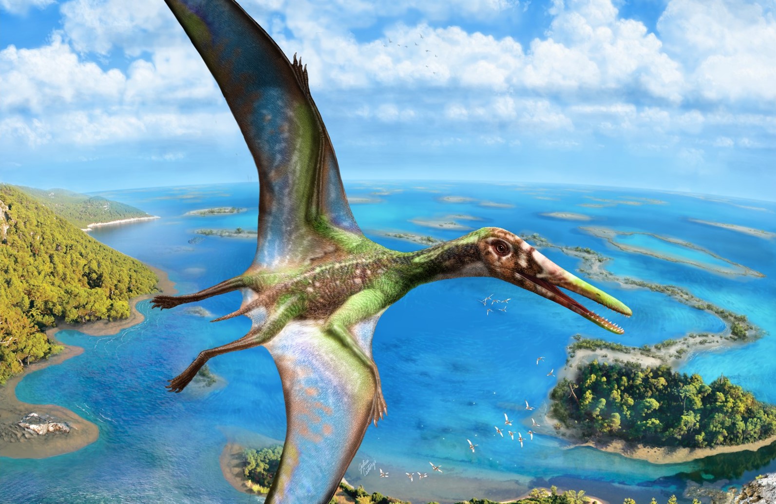
One of the studies involves a 150-million-year-old small-sized pterosaur with a wing span of approximately 40 cm, once believed to be incapable of taking off from water. The new study shows that it actually did possess the ability to overcome the water’s high surface tension and launch itself using both its feet and wings. This was during the Late Jurassic, 30 million years earlier than previous studies had found. The team used soft tissue data revealed in fossils through LSF to perform aerodynamic modelling showing that this pterosaur was capable of a “quadrupedal water launch”, similar to how ducks take off from water today. Dr Pittman says that LSF made it possible to reveal invisible details from the pterosaur’s wing membranes and webbed feet.
Another outcome was discovering a long umbilical scar in a 125-million-year-old Psittacosaurus dinosaur fossil that points to it being the equivalent a human belly button. This discovery sets a new record for the oldest belly button ever found. Dr. Pittman explained that dinosaurs did not have an umbilical cord because they laid eggs. Instead, the yolk sac of dinosaurs was directly attached to the body via a slit-like opening. When the egg hatched, this opening sealed up, leaving a distinctive long umbilical scar. LSF imaging identified the distinctive scales that surrounded the long scar in the specimen.


(Lower) LSF image of the whole Psittacosaurus specimen showing the location of the umbilical scar. (Image credit: Jagged Fang Designs; Bell et al. 2022.)
Fine soft tissue details in dinosaur fossils rarely preserve as they easily decay after death. With LSF, it turns out that many fossils have hidden soft tissues that are invisible under traditional white light. Looking at just the bones, you are limited when working out the origins of flight, so a big theme of Dr Pittman’s work is to gain more anatomical evidence through technology, which led him to co-developing LSF.

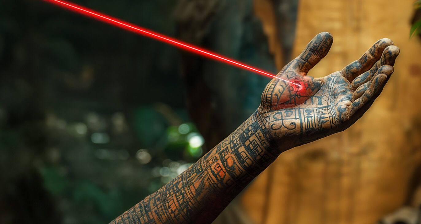
“Lasers have been around for years, UV (ultraviolet) lamps even longer. UV can make features of bank notes and fossils glow in the dark, but LSF is UV on steroids,” he explains. It involves scanning a high-power violet laser (near-UV) over the target object in a darkened room. “The laser causes the fossil to glow in the dark as it interacts with the minerals within the fossil. When it hits the minerals, it bounces around the atoms like a ping pong ball and how it does that is reflected in its new colour when it emerges. This gives a qualitative indication of the mineral’s composition. By scanning the laser across the specimen, a qualitative chemical map of it can be generated. This can reveal parts of the fossil you cannot normally see like skin and feather patterns. I have studied thousands and thousands of fossils with the laser, but when I make a discovery with LSF, I am always lost for words.”
Not all laser colours work well. Dr. Pittman has figured out over time that, for feathered dinosaurs including early birds, if he uses a violet laser, its wavelength “does a beautiful job” in revealing their once-invisible anatomy. Technology has advanced and costs have come down making violet lasers more affordable and he has revisited past work done previously with a UV lamp. “We are seeing more and more details as laser technology advances and we expect to see even further improvement in the future, which is hugely exciting,” says Dr. Pittman. “We have done it with feathered dinosaurs and pterosaurs, and more recently in archaeology.”
World’s first application of LSF on artefacts – ancient fingerprints revealed

Another paper just out last month led by Dr. Pittman and Kaye demonstrates this method on archaeological artefacts for the first time. They have had success in finding previously unseen details on well studied items from the Roman town of Verulamium (around AD50 – 450) in what is now St Albans in Southeast England. This includes, the 2000-year-old fingerprints of a workman who handled a vase while it was still damp and a painted symbol closely resembling the Greek letter gamma (Γ) on a bowl, images only possible with LSF. LSF imaging not only plays the role of a frontline archaeological identification tool but also ambitiously evaluates conservation by tracking if restoration works were done properly and were recorded.

Dr. Pittman has been interested in both palaeontology and archaeology from a young age and his enthusiasm for archaeology is expanding with more amazing details revealed through his laser technology. “Laser work is not my research completely, it is a means to an end, so I can answer the most interesting questions. As a follow-up to that, I am going to Belize this month to look at the Maya civilisation and hopefully we will make some discoveries there. The real benefit of doing both fields is cross pollination. I can take this technology across disciplines.”