


In recent years practitioners of minimally invasive surgery worldwide have been imagining a scene in which a robot stands by the operating table serving as a surgical assistant to the surgeon; altogether a process with fewer interruptions, fewer discomforts and fewer incision ports for the patient. Professor LI Zheng, an associate professor in the Department of Surgery, Faculty of Medicine at CUHK, has just taken a step towards making imagination reality. He and his team at the department’s Multi-scale Medical Robotics Centre have invented an intelligent magnetic anchored and guided endoscope (MAGS endoscope), which will give surgeons an unimpeded, uncluttered and uninterrupted view of what they are doing and all but eliminate the need for a human assistant.
AI assisted detection providing a hands-free view
The current endoscope used in chest and abdomen surgery is a thin metallic arm with a camera at its end, hand operated by a surgical assistant to follow the surgeon’s instruments as he or she works. This process places limits on the surgeon’s field of vision and, unfortunately, the endoscope and the instruments are competing for space in the operating cavity and sometimes clash. Professor Li has introduced AI and robotics to liberate the endoscope so that it can roam over the scene and provide multi-angle views automatically.

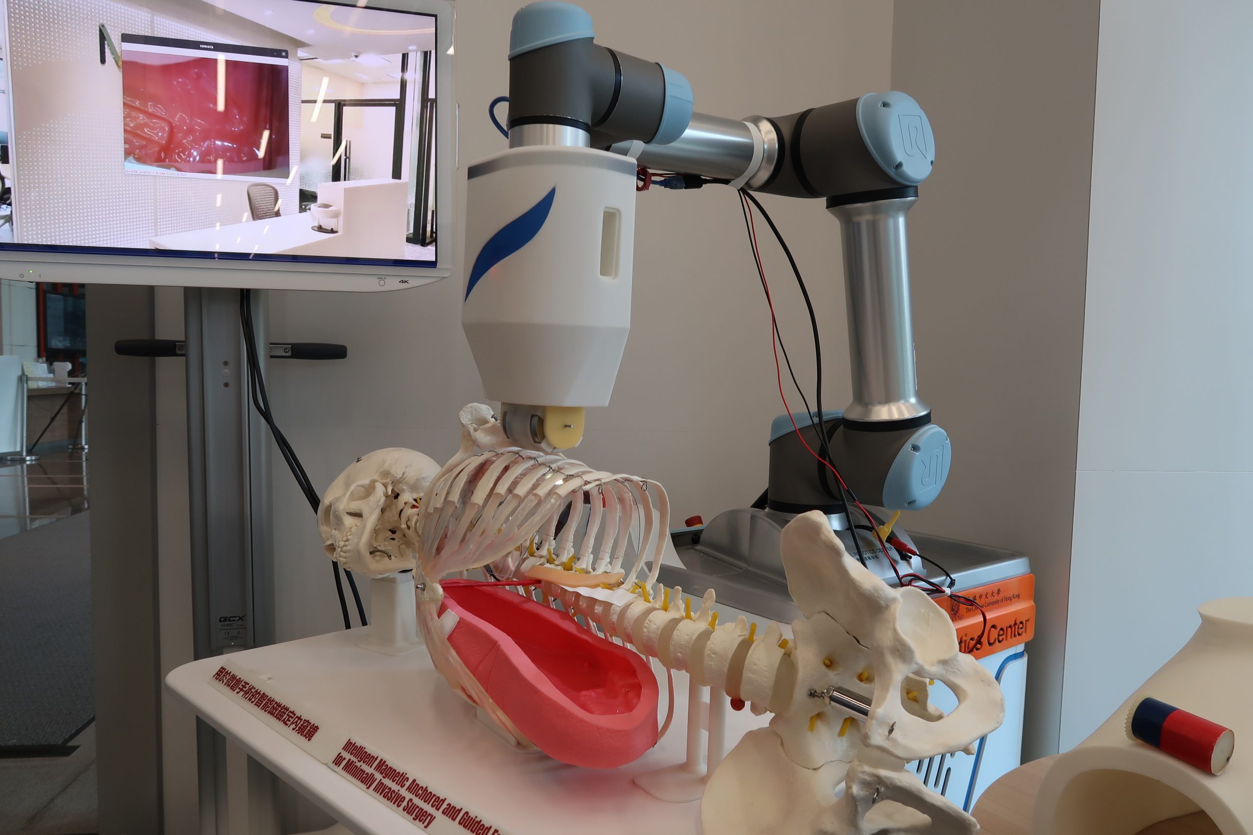
The MAGS endoscope can be inserted into the surgical cavity and track surgical instruments automatically. There are two main components. At the side of the operating table is a free standing robotic arm with a magnet at the end which rests on the outside of the patient’s chest. Inside the operating cavity is the endoscope in the form of a capsule with an LED light and magnets at each end. The endoscope is held to the inside of the chest wall by the magnetic pull of the external arm, observing the surgical scene from above.
The robotic control of the MAGS endoscope guides it to slide to different positions under the chest wall providing various perspectives with the camera. When the AI has identified the target, the tool designated by the surgeon, the visual servo control moves the camera to follow the target where it will work. “The motion relationships are mathematically pre-modeled by thoroughly training the AI,” explains Professor Li. “Intense training and rehearsal of the AI to recognise the target is essential. Surgeons can then work intuitively without worries about endoscope control.”
Self-cleaning function bringing a clear visualisation

This invention brings other bonuses. The conventional endoscope occupies too much port space. It requires a bigger incision or even an additional one, all of which means additional trauma to patients. The MAGS endoscope eliminates that, lessens the surgeon’s burden and smooths the surgical procedure by reducing instrument fencing. Most importantly, the camera in the magnetically controlled endoscope has been made self-cleaning. In surgery, the lens is frequently obscured by blood. The conventional endoscope has to be withdrawn from the cavity to be cleaned and the operation is halted. This can happen as often as twenty times during a procedure.
The MAGS endoscope has a soft 2mm rinsing tube connected to the camera. When needed, water is injected down the tube to flush the lens, followed by dried air. “In future, we could use AI to recognise blurring and automatically trigger an external mechanism,” hopes Professor Li.
The development of MAGS endoscope as a clinical product has inevitably faced difficulties. One of the challenges was to build a multi-expertise team with a balance of disciplines to develop all the technologies involved; for example, robotics, AI, magnetics and control and design. “On the other hand, the camera we are using and its imaging quality are also a concern. The more advanced the camera, the clearer the vision that is provided. The advancements in the medical camera industry will also benefit the MAGS endoscope in the long run,” explains Professor Li.
Enabling the future of surgery in chest and lung cavity

According to Professor Li, the magnetically guided endoscope was first conceptualised fourteen years ago. Since then, groups of scientists have been working on it but each with a different focus. “We started the project in 2015 and we are the leading group using robotics and AI for automatic MAGS view control,” he says.
The early models were designed around the abdominal cavity. However, the increase in incidences of lung cancer which took that disease to the top of the table of cancers in Hong Kong and the world made it clear that the current demand for MAGS endoscope is in chest surgery and the lung cavity. “This is what the present model and the team are working towards. It is still in development and we hope that the new version will go on to be tested in two to three years and that clinical trials will follow in three to four years and ultimately meet regulatory requirements,” says Professor Li.
This novel MAGS endoscope was awarded the Bronze Medal in the International Exhibition of Inventions Geneva 2021 and Gold of EMedIC Global 2019.