


Maths is often seen as esoteric. What little we know about maths may be memories of calculations, theorems, or equations we were given to tackle at school. In fact, maths is more involved in our lives in many ways, through computer graphics, animation or face detection. One of its most significant applications is medical imaging. Professor Ronald Lok Ming Lui from The Chinese University of Hong Kong (CUHK) has brought maths even closer to us by applying Computational Quasi-conformal geometry (CQC) to the analysis of medical imaging.
Professor Lui nearly left maths behind at school too. He was all set to apply to study medicine at university but he discovered that he was scared by the sight of blood. That profession’s loss was still medicine’s gain. After obtaining his PhD in Mathematics from the University of California, Los Angeles (UCLA) in 2008, and post-doctoral studies at the Harvard University under the supervision of Professor Shing Tung Yau, Professor Lui joined the Department of Mathematics at CUHK in 2010 with a profound commitment to developing CQC and applying it to medical imaging, particularly of the brain.
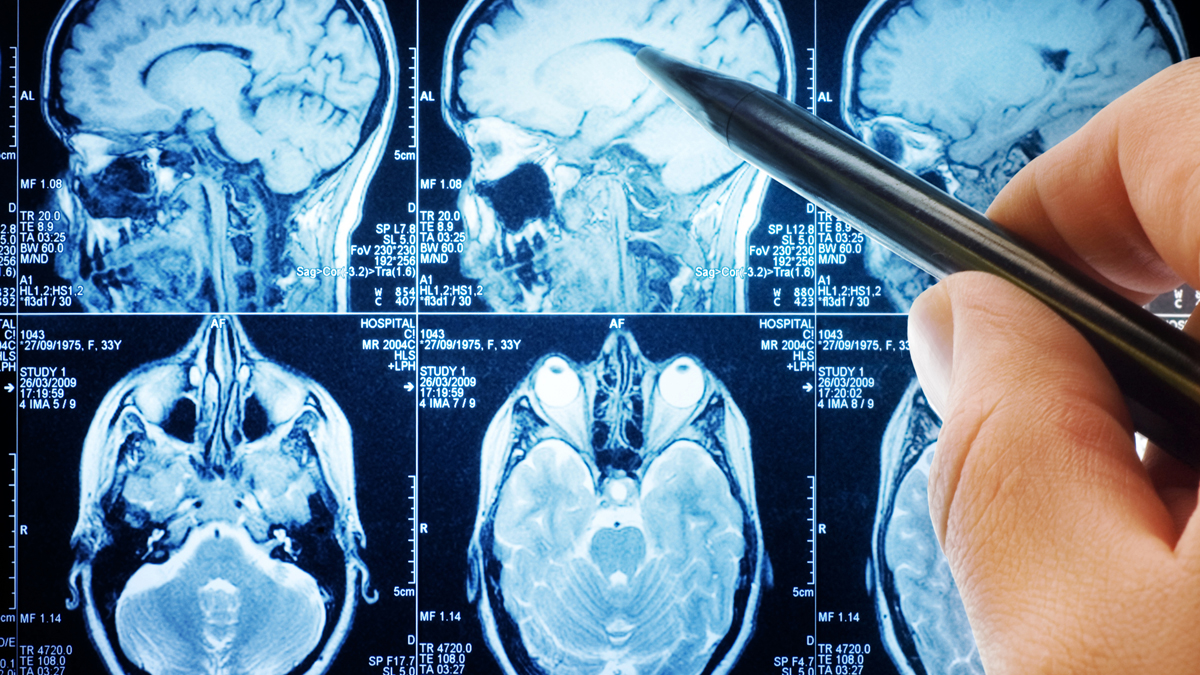
“You can create 3D images by using 3D camera and scan from different angles; you can even use a cellphone but medical imaging is totally different,” he says. “You take MRI scans, basically slices from different directions which give you a whole bunch of 2D images. But one thing is missing. You have lost all the geometry. When you look at the brain, there is a lot of geometry; ridges and depressions and this is very important for analysis. So is it possible for us to reconstruct this 3D surface?”

In his research, Professor Lui developed mathematical models using CQC. These models capture the boundaries of the object in each image. All these boundaries are combined to get a 3D optic. Accuracy in doing this is important or any analysis doctors derive from the image will be useless.
How do you actually construct the image? “The CQC image is made up of many triangles”, explains Professor Lui, “The mathematical formula that you use to extract the boundaries and put them together in a 3D optic creates these triangles which are a construct to deform and translate the information into a 3D form.”

An early collaboration with the Faculty of Medicine at CUHK (CU Medicine) came in 2011 when Professor Lui helped to define deficiencies in the vestibular system or sense of balance which was discovered to be the cause of spinal distortion in primary school age children. Little could be gained from an MRI scan of the inner ear, a complex interconnection of circles where the problem lay. However, CQC’s differential geometry analysed surfaces by numbers and gave an accurate description of shape and size. It was the first time this had been done anywhere in the world.

It was the brain though which drew most strongly on Professor Lui’s interest. “I like medical applications because I was thinking of being a medical doctor. Also my grandmother passed away from the complications caused by brain injury so that’s why I try to study it by maths.”
To Professor Lui, the brain is one of the most complicated items for which to develop a mathematical model. “There are lots of ridges and valleys in the brain and doctors can use the human eye and identify fingerprints on these features when they see them, which is amazing, but I thought to myself there has to be some mathematical equation by which we can map them so they can see them clearly. This led me to research into CQC and apply it to map the brain,” he says.


At the time he was doing his PhD at UCLA, he developed mathematical models which helped doctors to label features on the brain. Human brain mapping had just started and this labeling was done by hiring undergraduates. It took an hour per brain to label ten features, but a mathematical process developed by Professor Lui speeded that up.
Professor Lui began his research by focusing on conformal geometry, which is very specific. However, he realised that conformal is mathematically beautiful but not realistic. “If I want to study the difference between my face and your face, I try to deform my face to your face but a perfect conformal of the two faces does not exist. If you are looking at a surface that has become diseased, you want to see how its shape has changed over time and such a deformation is not conformal. But quasi-conformal geometry can describe change and deterioration. That’s why I switched to quasi-conformal.”
Together with his collaborators from various fields, Professor Lui’s group was the first to study CQC and its applications. CQC was a theory in the realm of pure mathematics, where you know it exists but cannot use it, so he studied how to develop a computational algorithm to compute it out. In 2010, he developed a formulation that could compute the QC map of a complicated brain in 20 minutes. “Doctors can maybe wait that long” he says, “but a computer cannot. Ten years later (in 2020) we had it down to 1 second.”
The brain surface obtained from the MRI scans is deformed in real time on the screen into a sphere which rotates, showing the whole topography from which the doctors can immediately identify fingerprints to work from. This year, the team has even speeded up the process to less than a second with the use of deep learning and AI. “This is very important because now we have big data. When I was a PhD student the data was limited to 50 to 100 brains. Now, we are talking about dealing with 1,000 to 2,000 brain surfaces.”
Considering that doctors are the end users, Professor Lui found that the design of a software interface for a CQC system which meets their needs is important. “We cannot assume doctors have a professional knowledge of mathematical algorithms. What we did is to communicate with them to develop a useable software interface by which doctors can obtain useful analysis from the system or 3D images, in a few steps. It’s a challenge of inter-disciplinary research,” he says.
In applications, Professor Lui has been in collaboration over his research with people around the world, including an Italian institution to realise 4D printing. Now he is working with CU Medicine to identify the risk of sleep apnea in children by using CQC and to identify and forecast the risk of dementia. Undoubtedly, Professor Lui’s work has made great improvements to the effectiveness of CQC and that, nowadays, is very much by making use of deep learning.